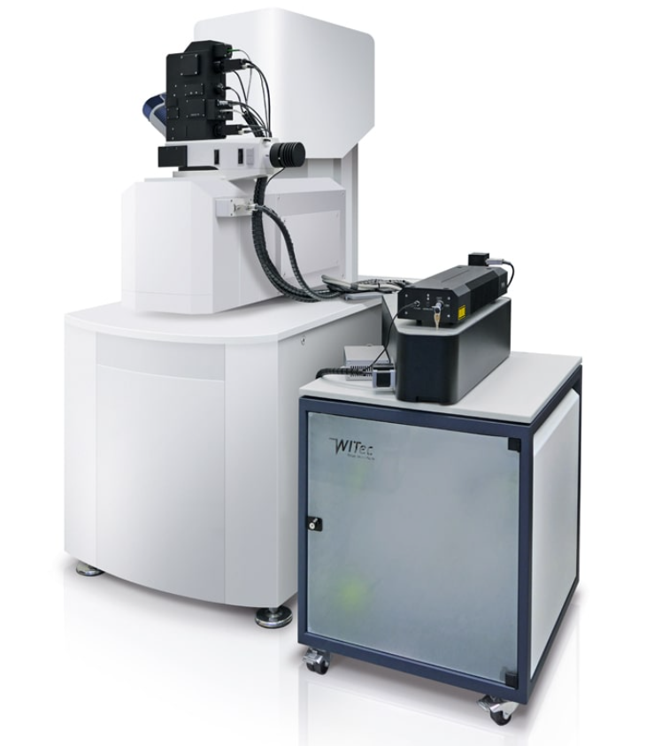
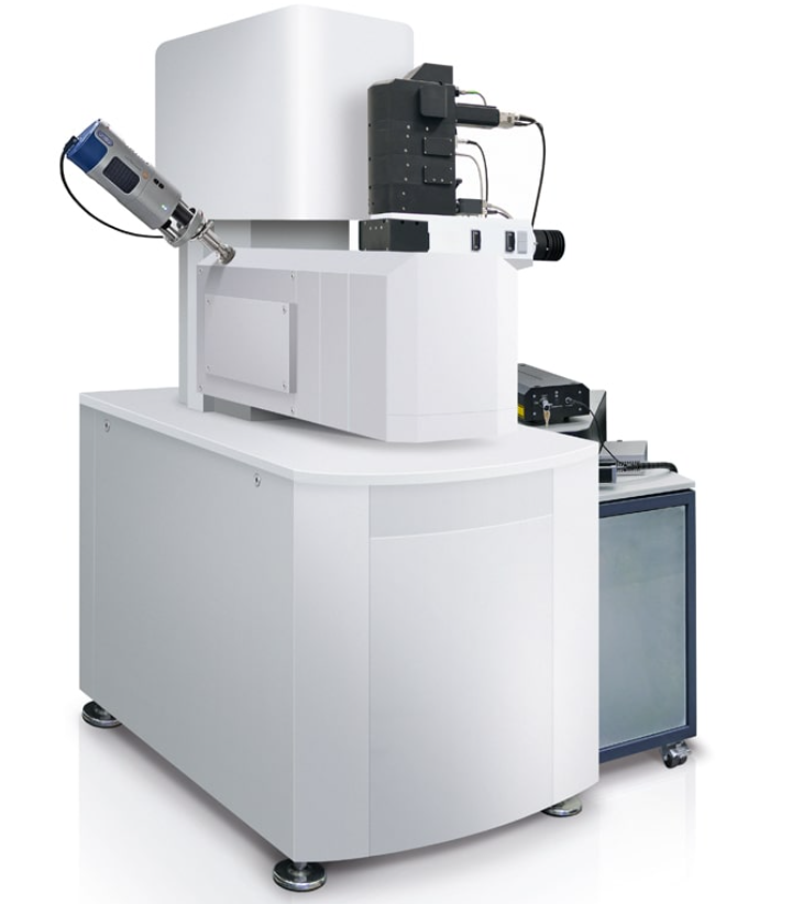
RAMAN-SEM MICROSCOPES (RISE) – OXFORD INSTRUMENTS (WITec)
THE RISE TECHNIQUE:
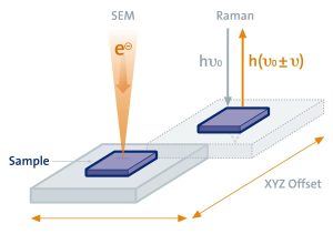 |
For RISE microscopy, samples are automatically transferred from one measuring position to the other within the vacuum chamber of the SEM, streamlining the workflow and drastically improving the instrument’s ease of use. |
RISE KEY FEATURES:
|
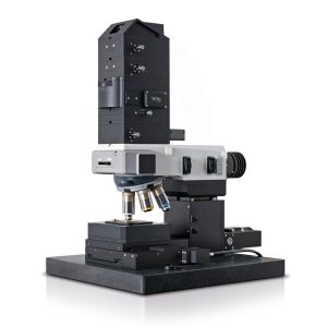
Scanning Electron Microscopy |
Confocal Raman Microscopy |
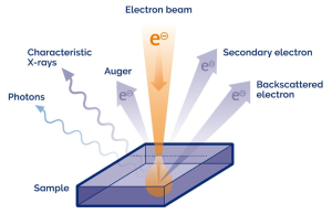
Scanning Electron Microscopy features a variety of analysis techniques. With RISE, the chemistry of molecular compounds can be imaged additionally. |
|
APPLICATION EXAMPLES:
 |
|
|
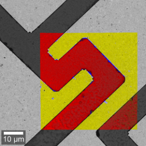
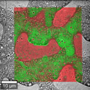
| Raman-SEM image overlay of a hamster brain tissue sample. Color-coded Raman image parameters: Green: White brain matter; Red: Gray brain matter; 100 µm x 100 µm, 300 x 300 pixels = 90,000 spectra, 50 ms integration time per spectrum. | Raman-SEM image overlay of a GaAs semiconductor sample. Color-coded Raman image parameters: Yellow: Gold substrate; Red: GaAs; Blue: Residues from production; 50 µm x 50 µm, 300 x 300 pixels = 90,000 spectra, 34 ms integartion time per spectrum. | Raman-SEM image overlay of a PMMA-PS polymer blend- Color-coded Raman image parameters: Green: Polystyrene; Red: Polymethyl methacrylate. |
|
|
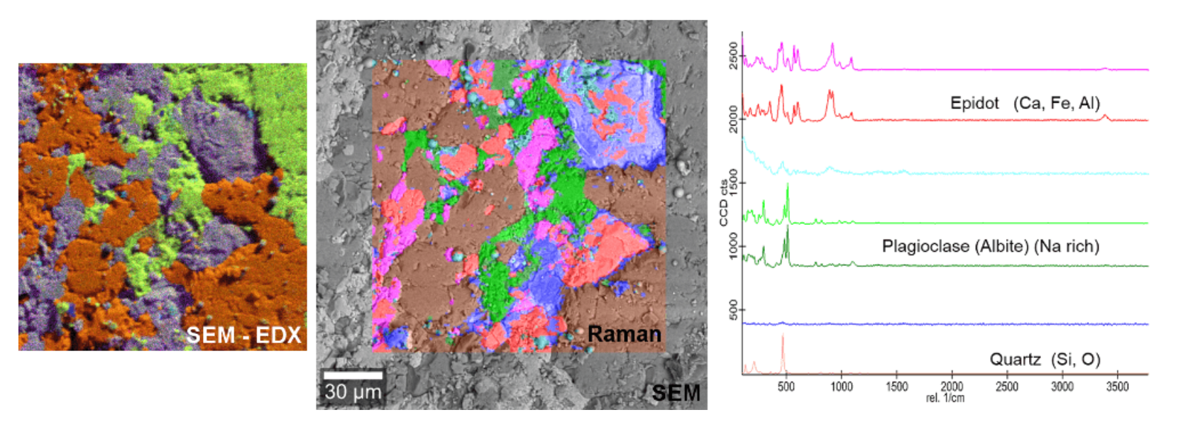
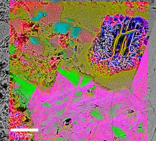
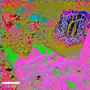
| RISE Microscopy and EDX analysis of a geological sample. Left: Overlaid SEM-EDX image: Three different element groups can be distinguished (Orange: Si, O; Purple: Si, Al, Fe, Ca; Green: Na). Middle: The Raman-SEM image overlay of the same sample area shows the distribution of the molecular compounds. Right: Corresponding Raman spectra. |